Brief description of the solution and the added value it delivers
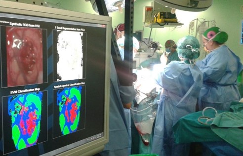
This hyperspectral image capture, processing and display system helps the surgeon to non-invasively delineate brain tumours during surgery. This technology displays the result in seconds, which means a reduction of up to 95% in the time taken compared to its direct competitor, intraoperative magnetic resonance imaging (iMRI). Furthermore, using this technology increases the precision with which the tumour can be located. This minimises the safety margin to be removed and improves the quality of life of patients, while avoiding possible relapses.
The prototype has been tested on more than 40 patients at two different hospitals, with satisfactory results.
Description of the technological basis
The proposed solution consists of a hyperspectral image capture, processing and display system that makes it possible to non-invasively distinguish between the different elements in the captured image. In brain cancer operations, it helps the surgeon to define the limits of the tumour efficiently and precisely.
Until now, identifying the edges of the tumour has relied on the surgeon’s experience, tests carried out prior to the operation and/or intraoperative magnetic resonance imaging (iMRI), which takes more than 45 minutes. Thanks to this technology, it is possible to work with information obtained and analysed in real time during the operation.
‘Precise delineation and graphic representation of brain tumours in a matter of seconds, using a non-invasive method’
Business needs / application
Health (detection of brain tumours)
-
Brain tumours are the cause of 2-3% of deaths worldwide.
-
Their tendency to infiltrate makes them difficult to demarcate.
-
The movement of the brain following craniotomy (brain shift) reduces the accuracy of the reference images.
-
The only alternative is intraoperative magnetic resonance imaging (iMRI), but this significantly increases the duration of the operation (more than 50 minutes per operation), with the consequent increase in the associated risks.
-
Surgeons determine the borders of the brain tumour based on their own experience, which is key to deciding on the safety margin to be removed.
-
A large safety margin may lead to serious consequences, significantly worsening the patient’s quality of life.
-
A small safety margin could leave remnants of the tumour, leading to its reappearance.
-
Minimising that margin may also help to reduce patients’ recovery time in hospital, as well as increasing surgeons’ productivity and, above all, reducing readmission, i.e. the likelihood that patients will have to return to the hospital with a problem resulting from the operation.
Competitive advantages
-
Hyperspectral image processing makes it possible to locate potential infiltration by the tumour.
-
Analysing images taken in the operating theatre counters the brain shift effect.
-
Those images are analysed in seconds – up to 95% faster than using iMRI.
-
The surgeon will have an additional source of information for demarcating the limits of the tumour and reducing the safety margin, improving the patient’s quality of life.
-
This tool will help to ensure that the tumour has been completely removed.
-
Each prototype costs around €250,000, which is 85% less than the alternative (iMRI).
References
-
This system has been developed thanks to the European project HELICoiD (HypErspectraL Imaging Cancer Detection, FP7-ICT-2013.9.2 [FET Open] 618080).
-
More than 80 images have been captured from more than 40 different patients. Those images have been validated by surgeons.
-
It has been tested at two different hospitals, one in Las Palmas de Gran Canaria and another in Southampton.
- Further development and optimisation through collaboration with Hospital 12 de Octubre, as part of the multi-year plan approved by the Community of Madrid.
Stage of development
-
Concept
-
Research
-
Lab prototype
-
Industrial prototype
-
Production
Contact
HYPER-EYE contact
Eduardo Juárez Martínez
CITSEM – UPM
e:
UPM contact
Innovation and Entrepreneurship Programmes
Technological Innovation Support Centre (CAIT) – UPM
e:


