BIOMEDICAL TECHNOLOGY CENTRE IMAGING SERVICE (BTC-IMS)
The BTC-IMS has several microscopes with very different imaging systems with the capacity and experience to look at cells, tissues and organs in physiological environments.
Description of the services offered
The BTC-IMS currently has 4 different microscopy systems:
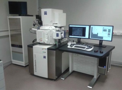
– Scanning electron microscope with an ion beam (FIB-SEM Zeiss Cross Beam 540)
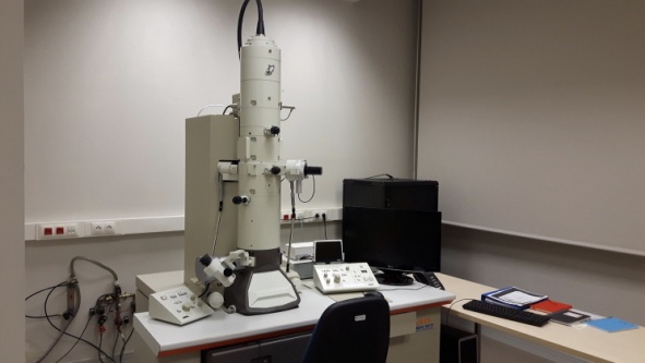
– Transmission electron microscope (TEM Jeol 1011)
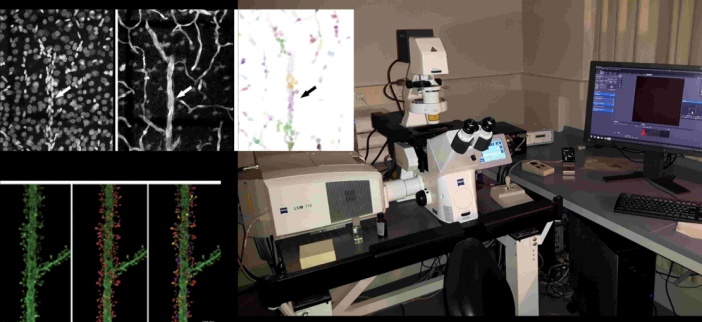
– Confocal microscope (MiC, Zeiss LSM 710)
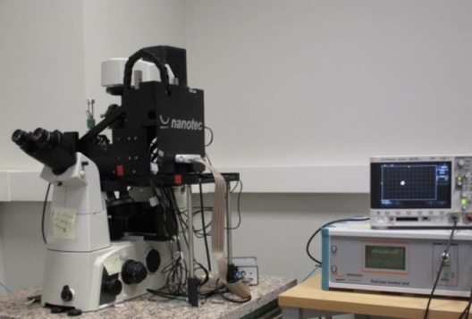
– Atomic force microscope (AFM, Nanotec Nanolife).
Needs requested and applications
The equipment at the BTC-IMS is available to make complete imaging studies of live specimens or fixed samples of tissues, organoids, 2D/3D cell cultures and various materials (polymers and gels, etc.). The equipment is, therefore, optimum for analysing the interaction of biological samples with structured nanomaterials and biomaterials. By way of example, the equipment is currently being used to carry out ex vivo studies of the cerebral cortex structure and analysis of plasticity in the cortex structure and cell activity in brains affected by different diseases, such as the study of murine models in Alzheimer’s or Parkinson’s disease.
Sector or area of application
Research in MEDICINE, BIOLOGY, MATERIALS SCIENCE, NANOMEDICINE, PHYSICS and CHEMISTRY
Differential skills
The innovative nature of this service lies in the opportunity to acquire very high resolution images in superficial and deep locations in biological samples, including tissues and 2D/3D cell cultures and their interaction with different biomaterials.
Previous references for provision of services
All the equipment is currently highly in demand for use in the field of Neuroscience and Bioengineering for reconstruction and replacement of damaged tissues and organs. At the moment, the equipment is particularly being used by R&D centre users with a high relevance in the field of neuroscience, and also in the field of biotechnology and biomedicine.
The Human Brain Project (HBP) and Cajal Blue Brain (CBB) project are being carried out at the BTC. In these projects, and with state-of-the-art anatomical techniques that make use of the BTC-IMS (MiC and FIB/SEM), it has been possible to construct a detailed map of the brain’s synaptic connections. In both cases, the equipment at the BTC-IMS is used with the aim of creating an anatomical model of mammals¿ brains at micro and nanometric scale.
Equipment description
The most important details about the equipment at the facility are as follows:
– FIB/SEM
Description: Zeiss Cross Beam 540 Field Emission Scanning Electron Microscope with Focussed Ion Beam.
Location: 30m2, Floor ¿1 within the Cajal Neural Circuits Laboratory area, 35A.S1.004.0, Edificio CTB, Montegancedo Campus UPM
Funding: 2016 Cajal Blue Brain Project (€800k), Human Brain Project (€130k)
Services offered: Acquisition of electron microscope sequential images.
For more information about requesting this services and its conditions of use, go to the BTC web site at the link: http://www.ctb.upm.es/core-facilities/
– TEM
Description: Jeol 1011 (100 Kv) transmission electron microscope equipped with a Gatan-Orius 11 Mpx camera.
Location: 30m2, Floor ¿2 within the Cajal Neural Circuits Laboratory area, 35A.S2.030.0, Edificio CTB, Montegancedo Campus UPM
Funding: 2010 Cajal Blue Brain Project (€350k)
Services offered: Image acquisition
For more information about requesting this services and its conditions of use, go to the BTC web site at the link: http://www.ctb.upm.es/core-facilities/
– MiC
Description: Zeiss LSM 710 Confocal Microscope equipped with: 5 lasers (405, 488, 543, 594, 633 nm), AOTF and AOBS systems and 2 GaAsP spectral detectors, on an inverted, motorised Zeiss Axio Observer Z1 microscope. Mercury vapour fluorescent lamp and filters for DAPI (445/50 nm), GFP (525/50) and Cy 3 (605/70).
Location: 30m2, Floor ¿1 within the Cajal Neural Circuits Laboratory area, 35A.S1.020.0, Edificio CTB, Montegancedo Campus UPM
Funding: 2010 Cajal Blue Brain Project (€250k)
Services offered: Image acquisition
For more information about requesting this services and its conditions of use, go to the BTC web site at the link: http://www.ctb.upm.es/core-facilities/
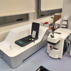
– AFM
Description: Nanotec Nanolife® atomic force microscope with 8 separate piezoelectrics, Dulcinea® control, WSxM software, liquid cell and XY positioner. AFM in contact mode (jumping, lithographic, dynamic (amplitude modulation, resonance frequency, frequency modulation); 2 (FZ), 3 (3D-Modes) and 4 dimensional spectroscopic measurements (General Spectroscopy Imaging including Force Volume); and long distance measurements using the retrace and plane scan methods. Automatic drift correction.
Location: 30m2, Floor ¿2 within the Biomaterials and Regenerative Engineering Laboratory area, 35A.S2.007.0, Edificio CTB, Montegancedo Campus UPM
Funding: 2010 Fundación Marcelino Botín Project (€220k)
Services offered: Image acquisition
For more information about requesting this services and its conditions of use, go to the BTC web site at the link: http://www.ctb.upm.es/core-facilities/
–
Ancillary equipment
The BTC-IMS has a full histology laboratory equipped with all the resources needed to process biological tissue.
Request for service
For more information about requesting this services and its conditions of use, go to the BTC web site at the link: http://www.ctb.upm.es/core-facilities/


