
Extracellular electrophysiology service
Extracellular electrophysiology service using the Multichannel Systems MEA2100 micro-electrode array system : In vitro recording and electrical stimulation of cell (neurones and cardiomyocytes) and tissue (brain) cultures.
Description of the services offered
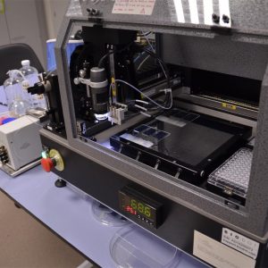
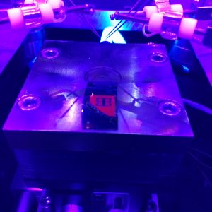
The MEA (micro-electrode array) system is equipment that enables simultaneous recording of the extracellular field potential generated by in vitro cell or tissue populations, making it possible to assess the physiological or pathological functioning of neurone or cardiac circuits with high spatial and temporal resolution. The MEA2100 system has an integrated amplifier and stimulus generator, temperature control and the option of real time detection and feedback. It has a sampling rate of up to 50 kHz per channel with 120 electrodes.
Needs requested and applications
The equipment offers essential, fundamental technology to understand learning and plasticity mechanisms, ageing processes and neurodegenerative diseases, and epilepsy and autism models, as well as assessing the behaviour of electrogenic cell cultures (neurones, cardiac, retina or muscle cells, and differentiated stem cells) under exposure to drugs or harmful substances.
Sector or area of application
Research
Differential skills
It involves many more benefits than other methods used for electrophysiological recording, such as the patch clamp or fluorescence. The MEA system takes non-invasive electrical measurements of a cell culture, without the need to use fluorescent proteins of calcium markers that may interfere with the cell model and confuse the results. Its non-invasive nature prevents mechanical damage to the cell membrane, unlike what happens with the patch clamp technique of glass electrode implants. Its high temporal resolution (over 50 kHz) exceeds visualisation using fluorescent proteins or calcium markers, which have a temporal resolution of under 100 Hz. It enables longitudinal studies to be carried out on the same cell population for hours, weeks and even months. Using the MEA is very easy. It simply involves cultivating the cell model on the electrode plates, putting the plate into the MEA system and taking the record. The data acquisition and analysis tools in the Multi Channel suite do the rest, with a simple interface for selecting filters, detecting spikes and electrical stimulation.
Previous references for provision of services
Where it is
The equipment is in the Biological Networks Laboratory at the Biomedical Technology Centre basement (35 A.S1.14). The centre has an animal facility and cell cultures unit, along with fluorescent, electronic and confocal microscopy services, meaning the MEA can be combined with image and optical stimulation techniques, making innovative use of optogenetics methodologies.